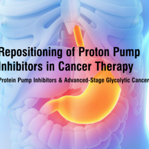
Repositioning of proton pump inhibitors in cancer therapy — download the full PPI from Cancer Chemother Pharmacol by Zhen-Ning Lui, Bing Tian, and Xiu-Li Guo.
Many of you may be aware that I have concentrated my cancer research on the glycolytic nature of many cancers. Cancers that are glycolytic, such as GBM and triple negative breast cancer, primarily rely on glycolysis for the generation of ATP, rather than mitochondrial ox-phos. The end product of glycolysis is pyruvate, which then will be converted into lactic acid. These glycolytic cancer cells must upregulate their mechanism(s) for effluxing lactic acid, or else they will succumb to necrosis or apoptosis, secondary to acidosis.
Any one or combination of 6 mechanisms may be upregulated, but commonly, the proton pump is one of the mechanisms upregulated.
A study was performed in 2012, in which adult females with metastatic breast cancer were randomly assigned to 3 arms: Arm A; Docetaxol 75 mg/m2 and cisplatin 75 mg/m2 on day 4, repeat q 21 days; Arm B; The same chemotherapy plus oral esomeprazole 80 mg bid on days 1-3, Arm C; The same as Arm B except esomeprazole 100 mg bid. Median progression free survival: Arm A (N = 33) 7.5 months; Arm B (N = 30) 10.9 months; Arm C (N= 31) 9.5 months. Among 17 patients with triple negative breast cancer this difference was bigger with median PFS of 9.5 and 3.3 months respectively.
The reason that the great difference was seen in the triple negative group, was that triple negative breast cancer is typically the most glycolytic of the breast cancers. Although the data indicates that long term use of proton pump inhibitors in the healthy population may lead to nutritional deficiencies, as well as altering the bacterial flora, the data suggests using PPIs may be a helpful adjunct in patients with advanced-stage glycolytic cancers.
healthyliving August 27th, 2019
Posted In: cancer care, Cancer Prevention
Tags: cancer, cancer care, glycolysis, glycolytic cancer, metastatic breast cancer, proton pump inhibitors

Testosterone Research Brings New Hope for Cancer Patients – an article from Science Daily.
This is certainly not the first article published on the benefits of anabolic steroids for cancer cachexia. Anabolic steroids, such as testosterone and nandrolone, not only reduce muscle wasting, they also increase erythropoietin release, thereby mitigating the anemia and fatigue associated with maximum tolerated dose regimens. This affords the patients less transfusions, as well as a decreased need for erythropoietin stimulating agents, which have shown to increase mortality in cancer patients.
Commonly recommended drugs for appetite stimulation, such as progestins, corticosteroids, and dronabinol, are minimally and transiently effective. In addition, there is no risk of promoting the aggressiveness of the cancer with anabolic steroids if the cancer does not possess androgen receptors. To the contrary, breast cancer survivors who received subcutaneous implants containing testosterone in combination with a low dose of anastrozole for relief of menopausal symptoms, have had no recurrence of cancer after eight years of testosterone therapy.
Bottom line: consider the use of anabolic steroids for cancer cachexia, as long as the cancer does not possess androgen receptors, such as prostate cancer and often triple negative breast cancer.
healthyliving June 20th, 2019
Posted In: cancer care
Tags: anabolic steroids, cancer, cancer cachexia, cancer care, nandrolone, testosterone

healthyliving May 14th, 2019
Posted In: cancer care, Cancer Prevention
Tags: cancer, cancer care, immunotherapy, oncolytic virus, OV, OV therapy, rigvir

“Circulating Tumor Cells Predictive of Adjuvant Radiotherapy Benefit in Early Breast Cancer” – an article from Cancer Therapy Advisor can be accessed here.
This article validates what I have been proposing for quite some time; we should be checking for circulating tumor cells (CTCs) in early stage cancer.
In the US, we have a test called CellSearch, to search for CTCs in breast, prostate, and colorectal cancer. This test is FDA approved to help physicians make clinical decisions in patients with metastatic breast, prostate, and colorectal cancer.
We know the greater the number of CTCs, in a patient with metastatic cancer, the worse the prognosis. This study found that approximately 20% of patients with early stage breast cancer had CTCs. This study also showed that more aggressive treatment, adding radiation therapy, in those with CTCs, improved survival.
One of my mantras has always been, “the practice of medicine lags far behind the science of medicine.” This scientific data can and should be applied to the practice immediately. On early stage cancers, we should be checking for CTCs. If we are dealing with breast, colorectal, or prostate cancer, we can use the CellSearch test for those in the U.S. If it is a different type of cancer, we will need to send the patients’ blood outside of the country (such as a lab in Germany), where they test 6 other genetic markers for CTCs. If the patient has early stage cancer and CTCs, in my opinion, the adjuvant or neoadjuvant treatment should be continued until the CTCs are zero.
healthyliving February 7th, 2019
Posted In: cancer care
Tags: breast cancer, cancer, cancer care, radiotherapy, tumor

“Wound-Healing in Mice Triggers Growth of Dormant Tumors” – an article from TheScientist can be accessed here.
Comments from Dr. Rosenberg on the article:
This is yet another article that suggests that operating on cancer spreads cancer. But this article is not referring to the seeding effect (dislodging cohesive cancer cells via biopsy or surgery, allowing them to disseminate); rather, it refers to an inflammatory or perhaps an immune response (or lack thereof) that allows cancer to spread, in response to surgery.
Unfortunately, at least for the near foreseeable future, we have to operate on cancer. Based on multiple articles, however, there is sufficient data to recommend starting NSAIDS several days prior to cancer surgery, administering ketorolac during surgery, and continuing NSAIDS for at least several days following surgery.
Previous studies have suggested an association between the use of NSAIDs and better outcome after mastectomy and lung surgery for cancer. In a retrospective analysis, investigators found an association between intraoperative NSAIDs use in conservative breast cancer surgery and breast cancer disease-free survival: https://academic.oup.com/bja/article/113/suppl_1/i82/307662. Although surgeons will tell patients to stop all medicines/supplements that can possibly interfere with clotting prior to surgery, there is sufficient data to indicate the opposite recommendation, specifically for NSAIDS.
healthyliving January 23rd, 2019
Posted In: cancer care
Tags: cancer care, dormant tumors, NSAID, NSAIDs, operate, operating, tumors

“Could Diet Adjustment Prevent Aggressive Prostate Cancer?” – an article from Cancer Therapy Advisor can be accessed here.
Comments from Dr. Rosenberg on the article:
Please do not use the ketogenic diet for all cancers! Prostate cancer has a different metabolic phenotype than the classic glycolytic cancers, where the “Warburg effect” applies.
Prostate cancer tends to increase their uptake of amino acids, such as glutamine and arginine, as well as fatty acids. Prostate cancer also increases expression of the enzyme fatty acid synthase, to help synthesize long-chain fatty acids. As I mentioned in prior issues, there is significant data to suggest that statins may inhibit prostate cancer. Because of prostate cancer’s reliance on lipids, the FDG-PET scan is not an ideal imaging study.
So what kind of diet should prostate cancer patients follow? Low fat diet, but keep in mind, that as the cancer progresses and mutates, some of the lesions may become glycolytic, requiring low carbohydrate intake as well.
healthyliving January 10th, 2019
Posted In: cancer care, Cancer Prevention, Healthy Lifestyle
Tags: cancer care, diet, diet adjustment, healthy diet, prevent prostate cancer, prevention, prostate cancer

Here is another publication on the use of low dose chemotherapy, but instead of being delivered in a metronomic fashion, one of the chemotherapy drugs was delivered in a continuous fashion. You can read the article on the National Institutes of Health website, by clicking the title below.
As I have previously mentioned, the longer the interval between chemotherapy dosing, the more likely cancer resistance will ensue.
In the current work, 1-year survival was 82%, versus 45% reported for FOLFIRINOX (standard of care), and median overall survival was 19 months, exceeding the 11.1 months for FOLFIRINOX. The authors conclude with the following, “In spite of the depressing statistics resulting from mostly negative trials, oncologists continue to accept the marginal benefits of currently sanctioned therapies for pancreatic cancer…There is no conclusive evidence that these treatment protocols are better than the 5FU-based regimens which were used in the 1980s and 1990s.”
Where and how did the concept of maximum tolerated dose chemotherapy (MTD) arise? MTD protocols were developed initially in the context of treating childhood leukemias and lymphomas; their extended clinical applications to the treatment of solid tumor malignancies were based largely on the therapeutic successes observed in the treatment of Hodgkin’s Disease and childhood ALL.
Therapeutic modeling in solid tumors was largely based on the growth parameters defined by the growth of leukemia cells. The primary therapeutic target in the leukemias/lymphomas is the abnormal cancer stem cell population of the bone marrow/lymph node. This target is more amenable to treatment with cytotoxic drugs that block cell cycle proliferation than are solid tumors, as the propagation of abnormally dividing cells comprises the primary cancer phenotype.
Solid tumor malignancies develop a phenotype that is a product not only of genetically induced cell cycle dysregulation, but also as a consequence of the abnormal microenvironment created by the tumor mass. The net result is a tumor whose proliferative capacity is generally restricted to its outer margins, thereby seriously limiting the potential efficacy of cancer drugs that target dividing cells.
Bottom line: We are still using chemotherapy dosing for solid tumors based on studies and trials treating leukemias/lymphomas, when in fact, these are two entirely different disease processes. It is time for a Wake-UP Call!
healthyliving October 1st, 2018
Posted In: cancer care
Tags: cancer care, chemotherapy, solid tumor, tumor

Cancer patients can work to understand and make healthy lifestyle decisions during their treatment. Adaptive and integrative cancer care practices have long advocated for integrating healthy choices about things like exercise and diet into a patient’s lifestyle.
The American Cancer Society recently shared this article by Elizabeth Mendez. The findings show that exercise may in fact, make tumors less aggressive and more likely to respond to treatment.
You can access that article here. It’s a good read for anyone faced with a cancer diagnosis, or working with cancer patients.
healthyliving May 16th, 2017
Posted In: cancer care, Cancer Prevention
Tags: cancer, cancer care, cancer prevention
Throughout the history of cancer research, in both the conventional and alternative cancer research realms, there has been a cause and effect relationship that has been largely ignored. The ability of a cell to divide, whether it be a malignant or non-malignant cell, is dependent on cell volume, as well as membrane potential. As you will learn, there is also a relationship between cell volume and membrane potential.
Cells that are cycling (dividing), progress through the following phases: G1 (Gap 1) – this is the
phase where the cell is preparing for the next phase, which is the S phase, or DNA Synthesis phase. Once DNA synthesis is complete, the cell enters the G2 phase (G 2), where it prepares to enter the final phase, called the M phase, or mitosis. During mitosis, the cell divides into 2 cells. The cell volume is at its smallest at G1, and gradually increases its volume until it reaches its largest volume in the M phase. This should be intuitive, because the cell must become large enough to divide and then support two cells.
Throughout the cell cycle, the cell is constantly monitoring the volume. If the cell does not reach the desired volumes, the cell will be unable to progress to the next phase. There is a G1/S transition “checkpoint,” which commonly causes the cell to arrest at this intermediate stage, if adequate volume is not reached. When a cell is arrested due to inadequate volume, there are two possible ensuing events: either the cell will leave the cycle and enter G0, and become a dormant, non-cycling cell, or the cell will be recognized as non-viable, and undergo mitochondrial-induced programmed cell death (apoptosis).
Interestingly, cell volume correlates with cell membrane electrical potential. Cells that are rapidly dividing are in the “depolarized” state; meaning they have less negatively charged particles in the intra-cellular space relative to the extra-cellular space. Cells that are not able to divide are “hyperpolarized;” meaning they have much greater amounts of negatively charged particles in the intra-cellular space relative to the extra-cellular space. Bottom line: Rapidly dividing cells have large volume and are depolarized, while non-dividing cells have small volume and are hyperpolarized.
healthyliving May 3rd, 2017
Posted In: Cancer Prevention
Tags: cancer, cancer care, cancer prevention, cell volume
Cell Volume, Cell Membrane Potential, and its Relationship with Cancer Proliferation
Throughout the history of cancer research, in both the conventional and alternative cancer research realms, there has been a cause and effect relationship that has been largely ignored. The ability of a cell to divide, whether it be a malignant or non-malignant cell, is dependent on cell volume, as well as membrane potential. As you will learn, there is also a relationship between cell volume and membrane potential.
Cells that are cycling (dividing), progress through the following phases: G1 (Gap 1) – this is the phase where the cell is preparing for the next phase, which is the S phase, or DNA Synthesis phase. Once DNA synthesis is complete, the cell enters the G2 phase (G 2), where it prepares to enter the final phase, called the M phase, or mitosis. During mitosis, the cell divides into 2 cells. The cell volume is at its smallest at G1, and gradually increases its volume until it reaches its largest volume in the M phase. This should be intuitive, because the cell must become large enough to divide and then support two cells.
Throughout the cell cycle, the cell is constantly monitoring the volume. If the cell does not reach the desired volumes, the cell will be unable to progress to the next phase. There is a G1/S transition “checkpoint,” which commonly causes the cell to arrest at this intermediate stage, if adequate volume is not reached. When a cell is arrested due to inadequate volume, there are two possible ensuing events: either the cell will leave the cycle and enter G0, and become a dormant, non-cycling cell, or the cell will be recognized as non-viable, and undergo mitochondrial-induced programmed cell death (apoptosis).
Interestingly, cell volume correlates with cell membrane electrical potential. Cells that are rapidly dividing are in the “depolarized” state; meaning they have less negatively charged particles in the intra-cellular space relative to the extra-cellular space. Cells that are not able to divide are “hyperpolarized;” meaning they have much greater amounts of negatively charged particles in the intra-cellular space relative to the extra-cellular space. Bottom line: Rapidly dividing cells have large volume and are depolarized, while non-dividing cells have small volume and are hyperpolarized.
How Can We Mechanically Slow Cancer cell Proliferation?
Hyperpolarizing cells would stop proliferation, but it would be difficult logistically to preferentially hyperpolarize cancer cells. The data, however, clearly supports that cell swelling promotes cellular proliferation, while cell shrinkage, inhibits cellular proliferation. So… how can we shrink cancer cells?
First we need to understand how cancer cells swell.
How Can We Osmotically Shrink Cancer Cells?
If we keep albumin and sodium levels high enough in the bloodstream, the oncotic/osmotic pressures will be elevated enough to keep water and sodium from escaping. It has been demonstrated both in-vitro and in-vivo, that exposing cells (malignant and non-malignant) to a hypertonic medium (high osmotic pressure medium) stops cell division. Patients with cancer can be treated with regular infusions of albumin, and sodium as needed, to keep intra-vascular oncotic/osmotic pressures at the critical level, to prevent cancer cell proliferation.
healthyliving April 4th, 2017
Posted In: cancer care
Tags: albumin, cancer care, shrink cancer cells

2017 Copyright Healthy Living Group. All Rights Reserved
Dr. Mark Rosenberg received his doctorate from Georgetown University School of Medicine in 1988 and has been involved with drug research since 1991.